Spatial Biology: Key Questions for the Future

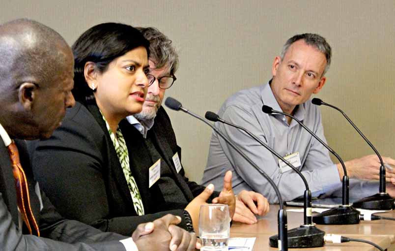
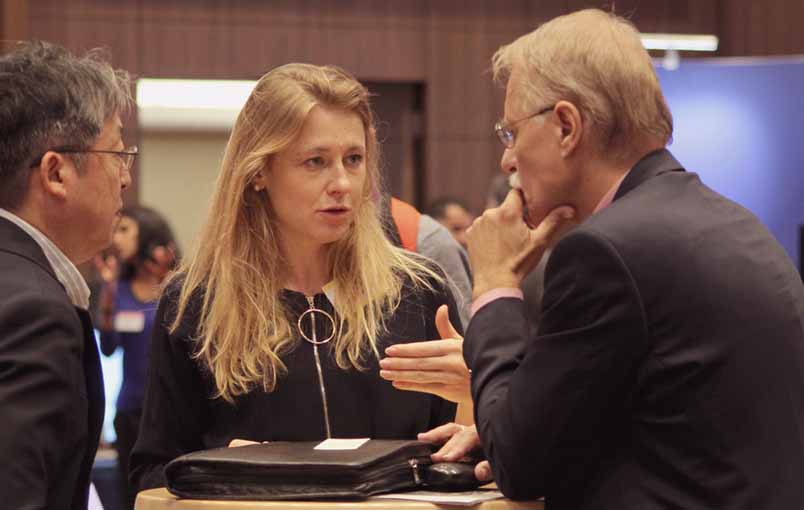
Our February discussion group brought together over 40 people working on spatial transcriptomics, technology advancements and data analysis. To facilitate such a large group a pre-session survey was sent out, asking attendees what topics they wished to focus on. The results proved to be very interesting and highlighted areas including single cell resolution, spatial analysis in oncology, and understanding the tumour microenvironment which were of particular interest to attendees.

This month’s group was led by Raffaele Calogero, Professor of Bioinformatics and Genomics Unit at University of Turin. Between 2002-2010 he organized, in collaboration with Affymetrix, training courses for biologists on microarray data analysis. Since 2010, he is involved in bioinformatics training at EMBL (Heidelberg). He runs courses on NGS data analysis in collaboration with Illumina in Italy, and in collaboration with DUKE-NUS in Singapore.
Spatial Transcriptomics to Single Cell Resolution
Currently, most spatial techniques do not provide insights at a single cell resolution. This is a concern, particularly in oncology, as many biological structures including tumours are made of heterogeneous cell clusters. One of the foremost goals of spatial transcriptomics is enabling single cell resolution on spatial platforms, which would eliminate the needs of running two independent experiments.
There are many technologies currently in development which are being designed with this goal in mind. Examples from the group included PhenoCycler-Fusion from Akoya Biosciences, which claims to be “the fastest spatial biology solution that enables ultrahigh-plex spatial phenotyping of whole slides at single-cell resolution”, and upcoming platform updates from Visium and 10x Genomics.
Key Question: “Do we need single cell resolution?”
“To study the tumour microenvironment, at least at the oncological level, that is something that you need. You need to be sure that those cells do not belong to the tumour itself. What we do in in our lab, if budget is sufficient, is run a single cell experiment plus the spatial. The problem is always the price of the overall experiment.“
Tissue Preparation and Reproducibility
Preparing tissue for spatial transcriptomic analysis is key to reproducibility and accurate results but remains challenging and expensive. Xiaoxia Liu et al (2022), conducted a study on tissue preparation for spatial analysis. They found that out of 125 samples from 57 pairs of lung cancer, paired‐cancer and normal tissues from 54 samples were suitable for spatial transcriptomes. Out of these, 43% were successfully prepared for analysis.
Key Question: "I'm wondering, since you're from a cancer field, how reproducible are your results? I'm from a neuroscience field and in my case, it's very reproducible, but it is heavily dependent on where the tissue is from and how it was sliced because if you're only 10 microns off, it results in an entirely different result."
"I would say that quality and reproducibility depend on the quality of the RNA you're working with. I like the idea of working on snap frozen samples, because the quality of the RNA is higher with respect the one observed on FFPE samples. Visium for FFPE technology is now on the market, and it seems to be quite robust, although the results are dependent on the shelf storage time of the samples. It is important to remember that the RNA quality of FFPE slides degrades progressively over the years. Definitively, experimental reproducibility is firmly linked to the quality of the of the input RNA."
Spatial Transcriptomics: Immaturity and Data Analysis
Spatial transcriptomics is a relatively new discipline, and it is generally agreed that current methods and tools are still developing. Raffaele explained that “spatial transcriptomics is not yet mature, in the sense that we have a lot of platforms. We’re more or less in the same situation that we had few years ago within the single cell RNAseq framework, when a plethora of different platforms was placed on the market. Today, only few of the initial platforms survived and now we have few clear market leaders. On these “survived” platforms are run most of the single cell experiments. The present problem for spatial transcriptomics is that if you decide to buy a specific platform today, and there are a lot of them now on the market, in a few years that investment may not pay out, because what you bought is no longer upgradable or supported.”
Another challenge is analysing the large volumes of data gathered from spatial experiments, particularly when combined with other data such as single cell omics. Current spatial transcriptomic techniques also face the issue of unwanted technical artefacts, which are not yet well characterised.
Key Question: “I think there is also immaturity of the spatial side when it comes to data and analysis. I've seen papers coming up with that are using the Google browser, I've seen people are using R and so on. So, what is everybody basically doing and kind of the immaturity of the domain as well?”
“What we do, in our lab, is picking the best of what is available within the open-source community. If we use clustering as an example, I can say that that most used tool by us is Seurat tool. We tested many clustering tools, and Seurat clustering implementation is the one performing better than the others. What we do normally, is running a conventional single cell clustering analysis on spatial RNAseq data and subsequently, overlaying the clustering results on the tissue slide. Although, it is a quite simple approach, we grab quite interesting info in that way, especially if we integrate spatial data with conventional single cell RNAseq.”
Final Thoughts & Conclusion
At Oxford Global, we couldn’t be more pleased with the turnout and feedback from this discussion. This group was one of our most popular sessions so far and provided the perfect setting for exchanging ideas, sharing innovations and discussing the ever-evolving spatial transcriptomics landscape. If you would like to join one of our future groups, consider becoming an omics community member. Future topics include single-cell analysis of the tumour microenvironment, target identification and single-cell proteomics.

