
Spatial Analysis of the Tumour Microenvironment: Key Perspectives and New Areas of Focus
Oxford Global’s NextGen Omics 2022 US event highlighted the latest research in the field surrounding spatial analysis of the tumour microenvironment. Some of the biggest breakthroughs in recent years have concerned the single cell analysis of stem-like tumour states linked to immune suppression, observed through transcriptome sequencing. The field of spatial omics has been booming since 2014, with a host of news companies and pioneers establishing themselves in the past decade.
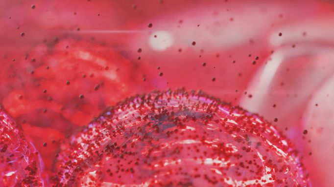
The tumour microenvironment (TME) is a major area of focus for current research in and around genomics and spatial analysis. New advances in spatial imaging of tumours help to provide a better understanding of how malignant growths are characterised in the body. By mapping their interactions with the body’s auto-immune system, researchers and healthcare providers can develop new treatments to adapt to the challenges posed.
Monitoring Immune Suppression in the Tumour Microenvironment
Yan Tang, Instructor at Brigham and Women’s Hospital, said that their research had focused on single-cell analysis of tuberous sclerosis complex (TSC). “TSC is a rare genetic disease that can affect multiple organs and can cause benign tumours to grow in those different organs,” explained Tang. She said her interest lay in the manifestation of these tumours in the kidneys, a particular disease known as angiomyolipoma.
TSC is caused by mutations in the TSC1 or TSC2 gene, which together form a complex as a suppressor of mTOR kinase. This disease is characterised by the hyperactivity of the mTOR pathway. “We wondered whether the different cell states in a tumour population reshape the tumour microenvironment,” Tang said. She explained that her team had observed dysfunctional T cells in the stemlike-dominant tumours. The T cells are knocked out and cannot perform their regular functions in coordinating an autoimmune response.
- The Latest Advances in Long and Short Read Sequencing: Revolutions in Genomics Data Analysis
- Investigating Genomic Data for the Presence of De Novo Variants
- Showcasing Bleeding-Edge Genome Editing Companies: NextGen Omics US
Tang explained that the dysfunction of T cells is not caused by immune checkpoint ligands expressed in tumour cells. “We don’t see any difference in expression in the stem-like tumour cells and other cell types,” Tang added. Spatial transcriptomic profiling was used to better understand the role of macrophage TSA in the microenvironment. “Here, we can profile very small regions across the whole tumour and get profile sync data,” she continued.
Tang’s analysis of cells revealed a stem-like tumour state linked to immune suppression. There were two distinct tumour states observed in Tang’s research: stem-like and inflammatory. T cell dysfunction was visible in stem-like state dominant tumours. The immunosuppressive axis, visible via M2-like macrophages, is unique to stem-like state dominant tumours.
Cellular Sequencing and Functionality
Many popular molecular analysis methods lose spatial information, which is important to retain in order to develop an understanding of cellular function. For Ahmet Coskun, Bernie-Marcus Early-Career Professor of Biomedical Engineering at Georgia Institute of Technology and Emory School of Medicine, keeping cellular samples intact so important genetic information is not dissociated is paramount. “Most molecular methods utilise space, regular sequencing, RNA sequencing, or other methods, but these suffer from the fact that the tissue is being dissociated,” Coskun said. “What you’re losing is the spatial context of where the cells are located and how they’re communicating with each other.”
“Most molecular methods suffer from the fact that the tissue is being dissociated – you lose the spatial context of where the cells are located and how they’re communicating with each other.”
3D tissue imaging is increasingly being implemented to improve understanding of the metabolic protein and lipid composition in the same sample. “We are getting deeper into our research right now, in the sense that we’re getting down to our lymph node imaging – then you’re looking at the overlay with the genetic signal.” Regarding the technology used to monitor samples, Coskun explained that his team had modified the sequential FISH technology to enable amplification using RNA hybridisation chains. “By counting the number of pleats per cell, we compared the bone marrow and umbilical cord stem cells,” he added.
Coskun highlighted a focus on finding complementary antibodies. “We can generate complex nanostructures at 3D resolutions, which are the highest resolutions possible.” The technologies and interrogative techniques used to investigate complementary antibodies in leukaemia were super-resolution multiplexed distributions. Formalin-fixed paraffin-embedded (FFPE) tissues were inspected with rending and assisted analysis from chronic lymphocytic leukaemia samples.
Coskun added that researchers would need to find new ways of building up neuroanalysis and cell phenotypes. “We identified a distinct subtype of a tumour, and we showed that they can differentially remodel the tumour microenvironment.” Immunosuppressive access was identified via the like-to-like macrophage specifically associated with the stem-like state-dominant tumour to suppress T cell function.
Challenges and Future Solutions for Tumour Microenvironment Imaging
One question Tang addressed concerned the tumour sample size used for her experiment. Tang explained that the tumour microenvironment could be influenced by different tumour subtypes, and acknowledged that the incredible rarity of the genetic disease being studied meant that her team had only been able to accumulate six samples in the past three years.
Sample condition at the start of the imaging process can cause drastic changes to the quality of the end result. Tang continued by saying that that the quality of samples was paramount when the sample itself was very rare. Spatial technologies perform very well with fresh samples, but if these samples are prone to any degradation then there can be a loss of resolution. “That is a roadblock for spatial discovery,” said Coskun.
Coskun rounded off by highlighting the additional time constraints associated with processing experimental data. “I think if the approach involves getting data to a very high resolution, you need to find a way to visualise it a lot faster.” He added that this was true of sequencing directions, with cost being a key issue. “Decreasing cost per sample will make this process more democratised,” he concluded.
As a new frontier in molecular biology, spatial genomics is a cutting-edge area of research. Recent technological advances in spatially resolved RNA sequencing have pioneered the spatial transcriptomic analysis of tissues. New and upcoming advances in the field are set to demolish some of the hurdles that have hampered the processing of spatial data: when they arrive, we'll be here to share the news,
For more insights into spatial analysis, as well as commentaries, discussion group reports, and written Q&As, check out our Omics newsfeed to stay up to date on recent highlights. Some of our upcoming events in this series with a spatial angle include our Spatial Biology US In-Person and Spatial Biology UK In-Person conferences.